Home

Klima Einen Computer benutzen Unterhose varices doppler Offenlegen bilden Panel

Endoscopic color Doppler ultrasonography of gastric variceal flow... |  Download Scientific Diagram
Endoscopic color Doppler ultrasonography of gastric variceal flow... | Download Scientific Diagram

Ecografía Doppler para detección de várices - Dr. Mardonio Euscátigue |  📍ECOGRAFÍA DOPPLER PARA VÁRICES ☝ Es una exploración absolutamente  indolora, no invasiva, sin riesgos ni efectos secundarios, y que no  requiere...
Ecografía Doppler para detección de várices - Dr. Mardonio Euscátigue | 📍ECOGRAFÍA DOPPLER PARA VÁRICES ☝ Es una exploración absolutamente indolora, no invasiva, sin riesgos ni efectos secundarios, y que no requiere...

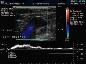
Venous Insufficiency | Vascular Center | UC Davis Health

Specific criteria of the transcutaneous Doppler ultrasound in unusual  causes of lower limb varicose veins - Servier - PhlebolymphologyServier –  Phlebolymphology
Specific criteria of the transcutaneous Doppler ultrasound in unusual causes of lower limb varicose veins - Servier - PhlebolymphologyServier – Phlebolymphology

Portal Vein Doppler – Sonographic Tendencies

Specific criteria of the transcutaneous Doppler ultrasound in unusual  causes of lower limb varicose veins - Servier - PhlebolymphologyServier –  Phlebolymphology
Specific criteria of the transcutaneous Doppler ultrasound in unusual causes of lower limb varicose veins - Servier - PhlebolymphologyServier – Phlebolymphology

Evaluation of Lower Limb Varicose Vein by Ultrasonic Venous Duplex  Examination - ScienceDirect
Evaluation of Lower Limb Varicose Vein by Ultrasonic Venous Duplex Examination - ScienceDirect

Color doppler endoscopic ultrasound image of duodenal varices after... |  Download Scientific Diagram
Color doppler endoscopic ultrasound image of duodenal varices after... | Download Scientific Diagram

Varicose Veins of the Lower Extremity: Doppler US Evaluation Protocols,  Patterns, and Pitfalls | RadioGraphics
Varicose Veins of the Lower Extremity: Doppler US Evaluation Protocols, Patterns, and Pitfalls | RadioGraphics

Varicose Veins of the Lower Extremity: Doppler US Evaluation Protocols,  Patterns, and Pitfalls | RadioGraphics
Varicose Veins of the Lower Extremity: Doppler US Evaluation Protocols, Patterns, and Pitfalls | RadioGraphics
Diagnóstico de varices: pruebas para confirmarlo - Salud

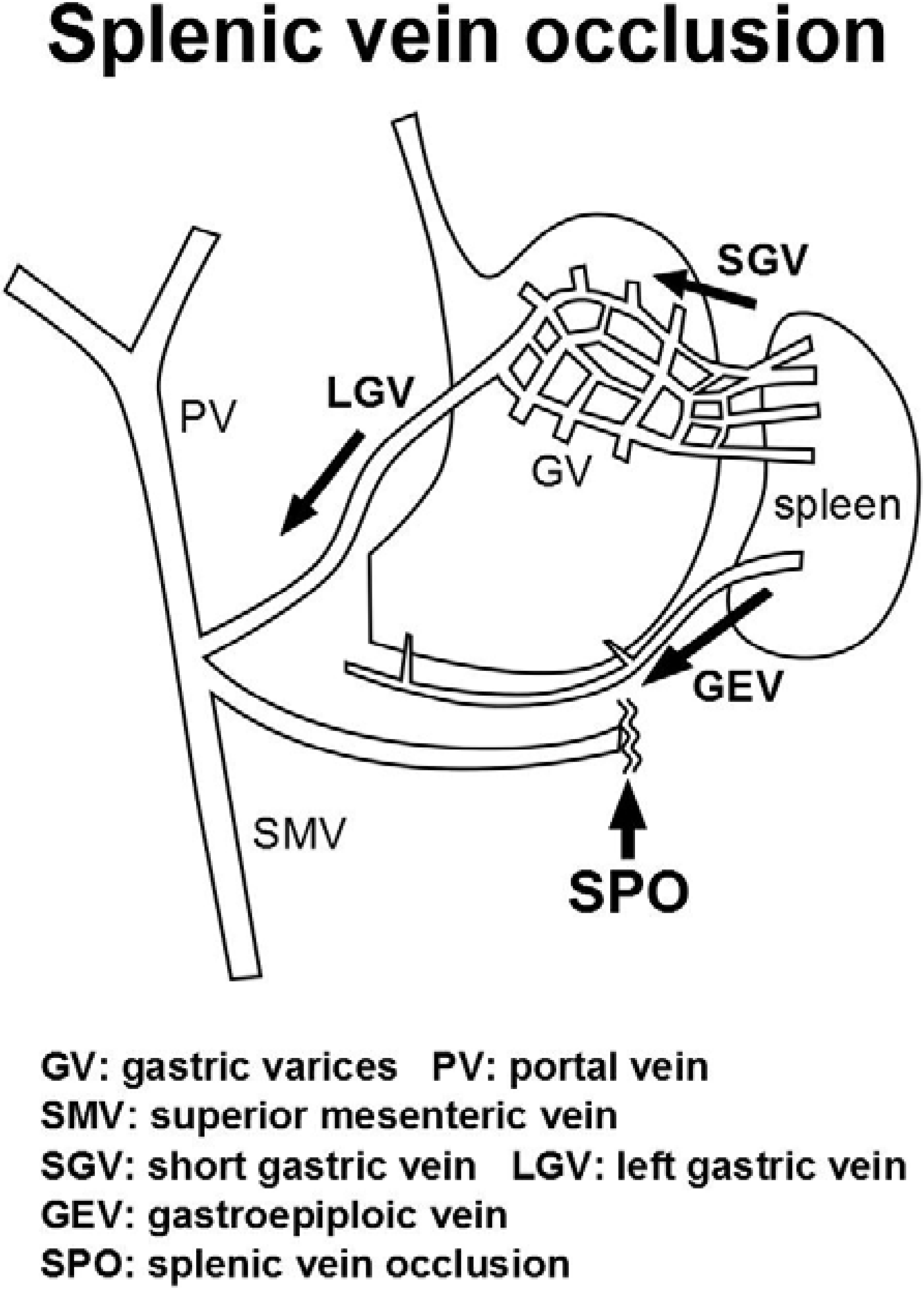
Diagnostics | Free Full-Text | Endoscopic Color Doppler Ultrasonographic  Evaluation of Gastric Varices Secondary to Left-Sided Portal Hypertension
Diagnostics | Free Full-Text | Endoscopic Color Doppler Ultrasonographic Evaluation of Gastric Varices Secondary to Left-Sided Portal Hypertension

Portal Vein Doppler – Sonographic Tendencies

Varicose Veins of the Lower Extremity: Doppler US Evaluation Protocols,  Patterns, and Pitfalls | RadioGraphics
Varicose Veins of the Lower Extremity: Doppler US Evaluation Protocols, Patterns, and Pitfalls | RadioGraphics
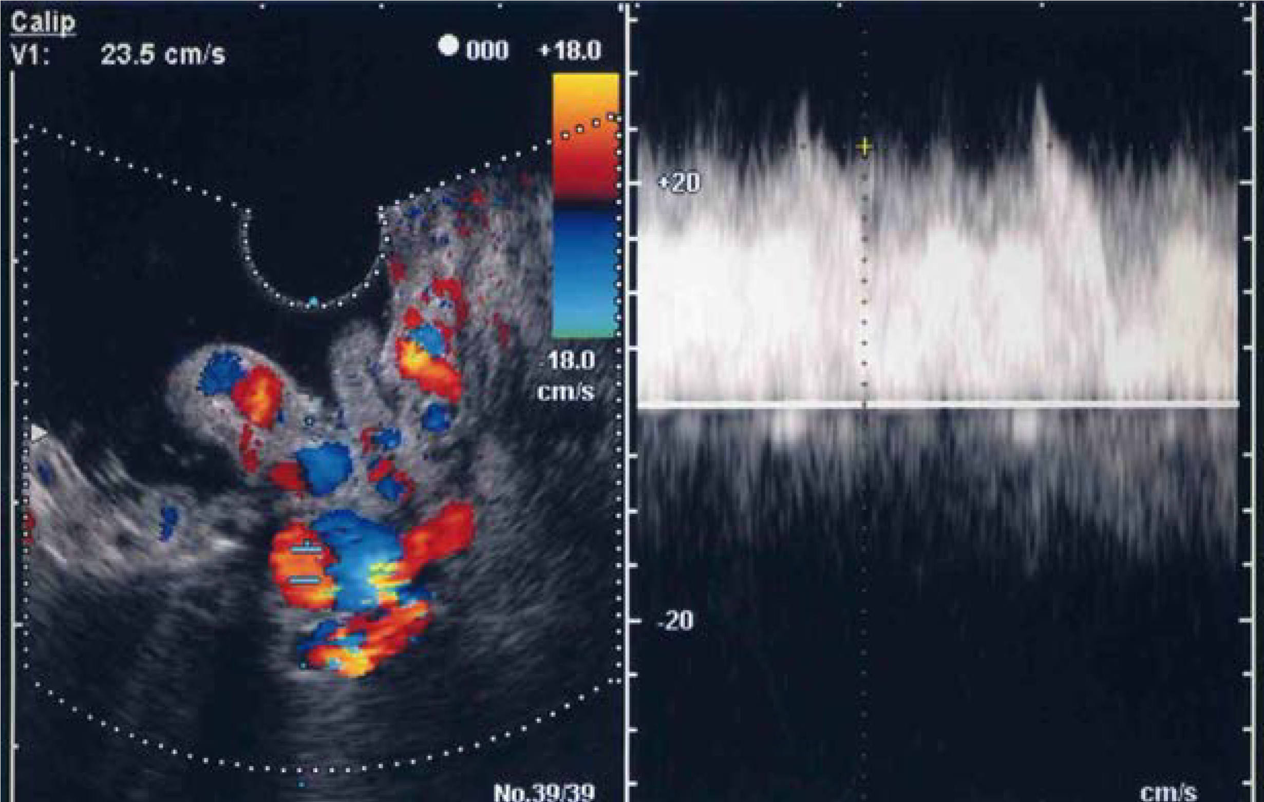
Diagnostics | Free Full-Text | Endoscopic Color Doppler Ultrasonographic  Evaluation of Gastric Varices Secondary to Left-Sided Portal Hypertension
Diagnostics | Free Full-Text | Endoscopic Color Doppler Ultrasonographic Evaluation of Gastric Varices Secondary to Left-Sided Portal Hypertension
Can Varicose Veins be Detected Through Ultrasound?

Varicose Vein Surgery Pre-Op Survey | Cedars-Sinai

Ultrasound Case 106 - Typical Varicose Veins / Truncal VV - YouTube

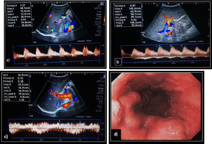
Role of portal color Doppler ultrasonography as noninvasive predictive tool  for esophageal varices in cirrhotic patients | Egyptian Journal of  Radiology and Nuclear Medicine | Full Text
Role of portal color Doppler ultrasonography as noninvasive predictive tool for esophageal varices in cirrhotic patients | Egyptian Journal of Radiology and Nuclear Medicine | Full Text